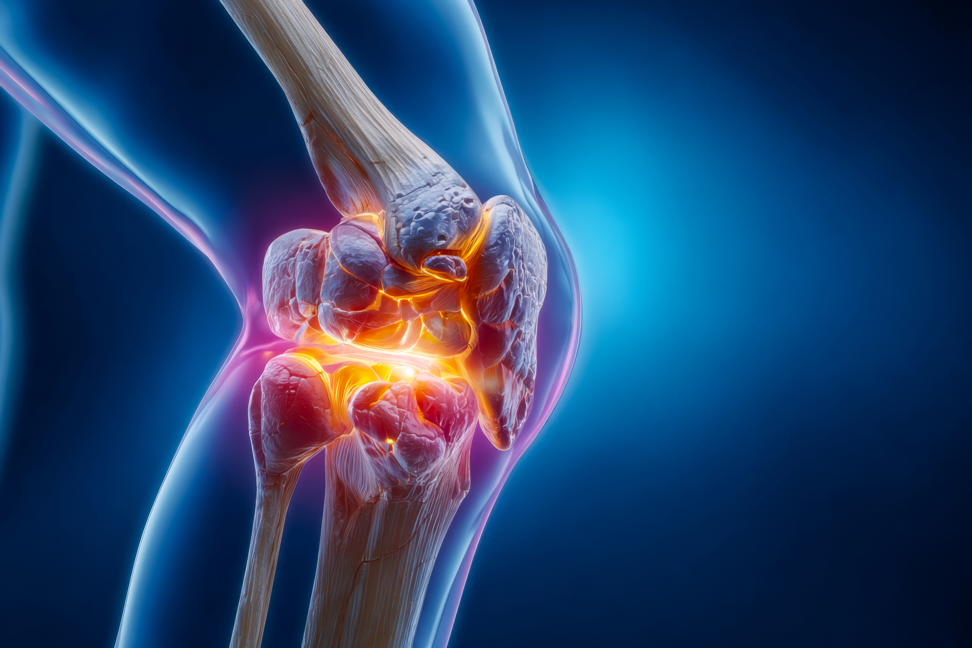
Do You Wake Up With Aching Joints or Stiffness That Makes It Hard to Move?
Maybe your knees hurt after a short walk, or your shoulders feel sore with every lift. These could be early signs of arthritis, a common condition that can slowly wear down your joints and affect your daily life.
Arthritis isn’t just about “getting older.” It can strike anyone, at any age, and often steals the joy from simple activities like climbing stairs, cooking, or even hugging your loved ones. But here’s the good news: modern imaging can help us see arthritis clearly and treat it early.
One of the best tools for this is the Arthrogram. This advanced imaging procedure lets doctors look deeper than a standard X-ray, capturing even the smallest changes in your joint’s soft tissues like cartilage and ligaments so you can finally get answers, relief, and a plan to protect your joints.
What Exactly Is an Arthrogram?
An arthrogram (pronounced ar-thro-gram) is a medical imaging test used to take detailed pictures of a joint after a small amount of contrast dye is placed into the space inside the joint. This dye helps highlight soft tissues such as ligaments, tendons, and cartilage, making them easier to see on imaging. Arthrograms are especially helpful when standard X-rays have not shown the full picture of what might be causing pain or limited movement.
Experts explain that arthrography is commonly used when doctors need a closer look at joint structures, particularly in areas such as the shoulder, knee, or hip, because these procedures can reveal subtle problems that standard images sometimes miss.
Why You Might Need an Arthrogram
At Monument Imaging, an arthrogram may be recommended when you have been experiencing:
- Persistent joint pain that won’t go away with rest or therapy
- Limited movement in your shoulder, hip, elbow, ankle, or knee
- Possible tears in soft tissues like ligaments or cartilage that weren’t clearly seen on other scans
An arthrogram can help your doctor answer important questions about your joint so that treatment, whether physical therapy, medication, or surgery, can be planned with confidence.
How the Imaging Works in Simple Terms
- Gentle Preparation – You’ll be made comfortable and your skin over the joint will be cleaned and numbed so you don’t feel the injection.
- Contrast Dye Injection – A small amount of safe dye is placed into the joint, which makes internal structures show up more clearly in the pictures.
- Imaging – After the contrast is in place, we use imaging such as X-rays, fluoroscopy, CT, or MRI to take clear pictures of the joint from several angles.
The whole process usually takes a short time, and most people can go home soon afterward. You may feel a bit of pressure or tenderness in the joint afterward, which is normal.
Why Arthrograms Are Still Valuable
Arthrograms have been used in musculoskeletal imaging for many years and remain an important tool when doctors want a detailed look inside a joint. The technique has evolved over time to complement advanced imaging such as MRI, especially when pinpointing issues inside a joint is critical for diagnosis and treatment planning.
In fact, arthrograms are often chosen in cases where doctors need extra detail to detect subtle tears, instability, or injury that might not show up on a standard scan.
We’re With You Every Step of the Way
At Monument Imaging & Diagnostic Center, we know that medical imaging can feel intimidating. That’s why our team focuses on compassion as much as technology, explaining each step, answering your questions, and treating you with care and respect.
We’re here to help you see what’s really going on inside your joint so you and your doctor can make confident decisions about your care.
📍 1201 Monument Road, Suite 101, Jacksonville, FL
📞 (904) 855-0700
🌐 www.monumentimaging.com
References:
- American Cancer Society. (2023). Brain and spinal cord tumors in adults.
- RadiologyInfo.org. (2023). CT scan of the brain.
- World Health Organization. (2021). Classification of tumors of the central nervous system (5th ed.).
- National Institute of Neurological Disorders and Stroke. (2022). Brain tumors information page.